I. What we know and what it can tell us about ourselves
Embryos are, or should be, familiar entities to people reading this blog, most likely biologists or scientists with an interest in Biology; but don’t take this knowledge for granted. Ask around what is an embryo and you will find difficulty in expressing an answer: a baby, a foetus, something small inside the womb of a pregnant woman, something to do with In Vitro Fertilization (IVF) are some answers I encountered when doing the exercise. “Embryo” is a word behind an entity which remains a big unknown. In the case of humans, probably the only embryos of interest to non-scientists, part of the mystery lies in their existence being hidden from view inside the maternal womb. Yes, we can get glimpses of embryos in the scans of our offspring where we want to see more than we can actually see, or in the apps that have become trendy to convey what is happening during a pregnancy, but this does not mean that, as a society, we know or understand what an embryo is. It is surprising that we are familiar with CRISPR, of course with genes, and also think we know a fair amount about the brain, to which we often liken computers, but, embryos?
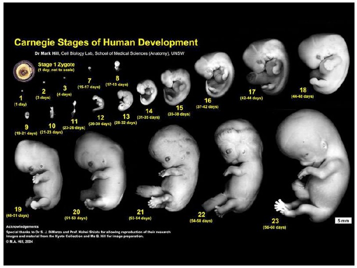
An embryo is not a foetus. In humans, an embryo refers to the period between the 3rd and 8th week after fertilization of the oocyte when the outline of what we shall become is laid down as the positioning of the primordia for organs and tissues along an orthogonal coordinate system: the head with the brain at one end; behind, the heart, muscles and bone, the gut and on the side small buds that will grow into limbs. Earlier, during the first three weeks, the fertilized egg multiplies and generates specialized cells that after a week surround a ball of cells that will become the embryo and provide a functional connection with the mother in the form of the placenta and a source of nourishment from the yolk sac. Then during week 3, the magic happens and through the process of gastrulation, the ball of cells is transformed into an outline of the organism, the embryo, which, during the next few weeks will organize, elaborate and connect the primordia, the seeds, for the different tissues and organs. A rudimentary heart is the first organ to emerge around the fourth week. From week 8 the embryo, now about 3 mm in length and with the design of the organism in place, is deemed to be a foetus and for the next 32 weeks will grow coordinating and fine tuning the development of the different organs and tissues. It is also around this time that a functional connection with the mother is initiated through the activity of the placenta. This, as all that follows, happens hidden from view, deep within the uterus but, nonetheless, we do know a fair amount of the aspect of early embryos and their transition to a foetus.
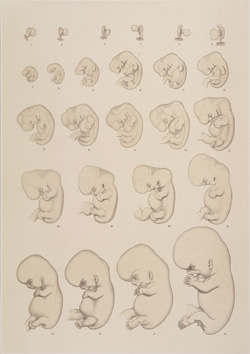
The observation and description of embryos of different animals was at the heart of Natural Sciences throughout the XIX century. Embryos from multiple species had been collected and their transformations over time recorded, analyzed, compared. Human embryos were scarce but some, and specially foetuses, could be found in private collections of medics or individuals with an inkling for the esoteric, in the forms of freaks (often ‘monsters’ associated with at the time unknown pathologies) more with the value of the outlandish than as elements of a Science to come (1). Many of the specimens of these collections can be seen today in anatomical museums around the world (see e.g ref. 2) but a systematic study of human embryos begins with Wilhelm His, who starting in the latter part of the century begins to fill the existing gap in knowledge between the development of humans and that of other animals (3). His creates a network of medics that provide him with embryos and foetuses that he then studies and arranges according to their degree of complexity. In 1880 he publishes the first timeline of human development with a special reference to the early stages (Fig.). The work was taken further by His’s disciple Franklin Mall who, after returning to the US and under the patronage of John Hopkins University and later the Carnegie Institution, creates the most complete collection of human embryos that exists to date (4). L. Morgan has told the story in a book that should be required reading for anybody with an interest in the human embryology (5). There are other collections around the world, notably a rather complete one in Kyoto (6), but none as complete and as intellectually satisfactory as the one that has become known as the Carnegie collection. The collection is still useful today.
Surprisingly, during this time, embryos, and human embryos in particular, feature in the public domain in ways that they have not done since. This is due to the efforts of the German biologist Ernst Haeckel. Enamoured of Darwin’s theory of Evolution, Haeckel had seen a way for a theory of his own with embryos at centre stage. Building on similarities between embryos of different vertebrates, described in particular by Karl Ernst von Baer earlier in the century, and blending them with the theory of Evolution, he posits the well-known mantra ‘ontogeny recapitulates phylogeny’. This was shorthand for a flawed grand view of Biology according to which organismal structural and functional complexity of supposedly higher order forms is achieved by recapitulating the development of simpler forms. Thus he suggested that in becoming a human being, we recapitulate (literally) the development of other animals. This was wrong before it was posed, as von Baer had already offered an alternative interpretation to those similarities, whereby during evolution the development of animals diverged and specialized from early common forms (7). However Haeckel, who on more than one occasion stretched the facts to fit his views, had panache, was a showman and reached the masses with popular books and talks about Evolution and his theories. The outcome was not only an idea of humans as a special breed but also of a flawed mechanism for how this was achieved. The history of this important episode in the history of Biology and its social and scientific implications are discussed in detail by N. Hopwood epic “Haeckel’s Embryos: Images, Evolution and Fraud “ (8). However, there was a message from these quibbles, one that in any event was there before Haeckel and that was used by Darwin in his great book, namely that embryos reveal our relationship to other animals. Before DNA, embryos had revealed our links to the rest of the natural world.
Throughout the first half of the XX century, the Carnegie collection, together with other smaller ones that emerge became a reference for texts of human embryology and medical studies. But the embryo, its transformations, remained hidden in the womb. Perhaps it was this mystery that created the furore of the article ‘drama of life before birth’ published by Life magazine in 1965 from the work of the photographer Lennart Nilsson. The photographs, later collected in a book “A Child is Born” portrayed the development of human embryos ‘inside the womb’; even though most of them are known to have been staged from abortions with consent. These pictures were a sensation and brought to light the embryo and the foetus in their natural environment but still left unanswered the mechanism behind the transformations.
The causal analysis of human development is the remit of Developmental Biology and involves experimentation: interference with the embryo. This analysis has its roots in the experiments of Wilhelm Roux and Hans Driesch in the late XIX century, emerges slowly during the XX century with the work of the Spemann school (9) and becomes a body of knowledge when it blends with genetics in the 1980s. The metamorphosis of embryology (descriptive) into developmental biology (analytical) is reflected in the emergence of journals like Developmental Biology in 1959 from the Society for Developmental Biology and in name changes like that of the Journal Embryology and Experimental Morphology (1953) into Development (1987) and the classic Wilhelm Roux Archives of Developmental Biology, founded by Wilhelm Roux to hail the emergence of experimental embryology in 1894, into Development Genes and Evolution (1996). Throughout this period developmental biology thrives with organisms like frogs, fish and chickens not to mention invertebrates, with Drosophila and Caenorhabditis elegans at the front. Mammals remain in the sidelines; their development inside the maternal womb creating a barrier. Nonetheless, slowly, the mouse begins to yield insights about the preimplantation stages (10) and through the systematic analysis of mutants in the 1990s, of the development of tissues and organs (11). The forces shaping the human embryo remain off bounds, restricted to the glimpses offered by the embryological collections, in particular the Carnegie and Kyoto ones.
Human developmental biology can be said to start with the work of Robert Edwards and Patrick Steptoe on In Vitro Fertilization (IVF). If you don’t know the story and think of IVF simply as a technique you’ll be surprised to learn how much fundamental biological research is behind this work (12, 13). The story is an impressive blend of medicine and biology, physiology and developmental biology, human tenacity and vision against funding adversity (14). It is this work that takes the early stages of human development out of the womb to allow their study. Having and growing a fertilized egg in a dish, as the early IVF practice requires, raised the question of how long would the fertilized egg survive and whether one could have an embryo on a dish. This led to a discussion of the associated ethical issues and questions associated with the potential of the experiment that, rightly, stalled research for a number of years. A committee convened around Mary Warnock made up of biologists, medical doctors, ethicists, religious leaders and lay people, to consider carefully the matter of when should one consider the start of human life. The outcome of these deliberations was the Warnock report (15) which in 1984 established a set of guidelines which have been followed by research with human embryos since. Importantly, amidst these guidelines is the 14 day rule that establishes this time as the beginning of the individual. It is this time that signals the start of gastrulation and was deemed to result in the emergence of the embryo as an individual entity.
As we move into the XXI century advances in developmental biology and genetics of different organisms are opening up new avenues for research and possibilities emerge to link genotypes and phenotypes. On our understanding of the emergence of vertebrate embryos, even of mice, advances come apace but the black box of human development, that period between weeks 3 and 8 remains hidden from us. Technical developments allow scientists to grow IVF discarded human embryos on a dish to the that 14 day barrier (16, 17) but it is not easy and there are technical challenges associated with these experiments. Notwithstanding this, we find ourselves in need to develop ways to gain insights into human development, to understand what drives those transformations that lie behind the images of the Carnegie and Kyoto collections, how come gastrulation takes one and a half days in the mouse and about three or four in the human embryo. We need to find ways that abide by existing ethical guidelines to address these questions so that:
- we learn how and why we are different from other organisms;
- we investigate the origin and causes of many pathologies and with this understanding explore ways to relieve them;
- we can understand, and perhaps ameliorate, the causes of early miscarriages.
Over the last few years, Embryonic Stem Cells have begun to open up avenues to address these questions by creating models of human development. These will be the subject of next week’s post.
References
- Leroy, AM (2005) Mutants. Harper Perenial
- Richardson, M.K. and Narraway, J. (1999) A treasure of comparative embryology Int. J. Dev. Biol. 591-602.
- Hopwood, N. (2000) Producing development: the anatomy of human embryos and the norms of Wilhelm His. Bulletin of the History of Medicine 74 (2000): 29–79
- Noe, A. (2004) The human embryo collection In “Centennial history of the Carnegie Institution of Washington pp 12-61. Ed J. Maienschein, M. Glitz and GE Allen.
- Morgan, L. (2009) Icons of life Univ. Calif. Press
- Yamaguchi, Y. and Yamada, S. (2018) The Kyoto collection of human embryos and fetuses: history and recent advancements in modern methods. Cells Tissues and Organs 205, 314-319.
- Gould, SJ (1977) Ontogeny and phylogeny. Belknap Press of Harvard University Press.
- Hopwood, N. (2015) Haeckel’s embryos: images, evolution and fraud. The University of Chicago Press.
- Hamburger, V. (1988) The heritage of experimental embryology. Oxford University Press.
- Alexander, H. (2001) A history of mammalian embryological research Int J Dev Biol 45, 457-467.
- Artz, K. (2012) Mammalian developmental genetics in the Twentieth century. Genetics 192, 1151-1163.
- Ball, O. (2012) Chapter 9 Opening the bottle in “Unnatural. The heretical idea of making people”. Vintage books London.
- Maienschein, J, (2014) Chapter 5 , The visible human embryo in “Embryos under the microscope”. Harvard University Press.
- Johnson, M., Franklin, S., Cottingham, M. and Hopwood, N. (2010) Why the Medical Research Council refused Robert Edwards and Patrick Steptoe support for research on human conception in 1971. Human reproduction 25, 2157-2174.
- Warnock, Mary. 1984. Report of the Committee of Inquiry into Human Fertilisation and Embryology. London: Her Majesty’s Stationery Office.
- Deglincerti, A. et al. (2016) Self-organization of the in vitro attached human embryo Nature 533, 251-254.
- Xiang, L. et al. (2019) A developmental landscape of 3D cultured human pregastrulation embryos. Nature 577, 537-542.